INVE600 — Inverted Cell Culture Microscope · Bright Field / Phase Contrast / Fluorescence
Inve600 inverted biological microscope Family
OVERVIEW
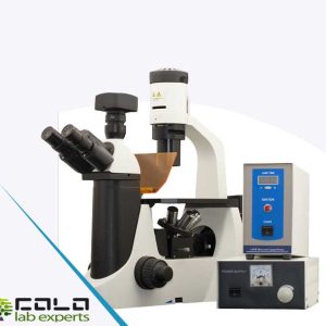
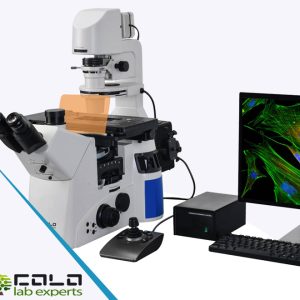
The INVE600 inverted microscope family is designed for laboratories that need a practical and scalable platform for cell culture observation, live cell work and advanced optical imaging workflows. The system combines a UIS2 infinity corrected optical system, inverted microscope construction, intelligent control functions, wide sample accessibility and flexible stage operation for modern laboratory use.
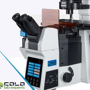
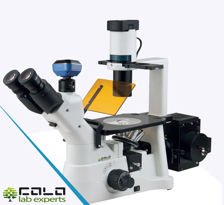
INVE600 is available in bright field, phase contrast and fluorescence configurations. With trinocular viewing, 3W S-LED 4000K Kohler illumination, quintuple nosepiece, LCD operating status display and application-oriented accessories, the platform supports routine microscopy, cell research, micromanipulation workflows and digital imaging integration.
Research-grade inverted microscope family with bright field, phase contrast and fluorescence-ready configurations, designed for cell culture workflows and digital imaging integration. INVE600 combines UIS2 optics, intelligent operation, practical stage handling and COLO.Science support for laboratories that need flexible everyday microscopy performance.COLO.Science · Practical and robust equipment for everyday laboratory workflows.
COLO.SCIENCE KNOWLEDGE
Practical insights for cell culture and inverted microscopy
Explore COLO.Science articles on inverted microscope selection, phase contrast workflows, fluorescence imaging, digital camera integration and practical cell observation routines.
Product Overview
INVE600 is an inverted microscope platform built for laboratories that require cell culture adaptability, large working distance, stable optical performance and configuration flexibility. The family includes INVE600B for bright field, INVE600T for phase contrast and INVE600F for fluorescence applications, making it suitable for laboratories that want one common platform across different microscopy tasks.
Configuration notice — delivery depends on selected model and options
INVE600 is a microscope family. The exact delivered configuration depends on whether the selected version is bright field, phase contrast or fluorescence, and on the selected optical accessories, filter modules, digital imaging adapter and workstation package. The official HTML/PDF technical specification defines the exact supply scope and available options. Specifications may change without notice.
Key Features
Inverted Platform
Bright Field / Phase / Fluorescence
3W S-LED 4000K
Kohler Illumination
Quintuple Nosepiece
Intelligent Operation
LCD Status Display
COLO Workstation
MicroscopyFlexible inverted microscopy for cell observation and laboratory imaging workflows
Cell Culture Ready
The inverted microscope format, removable condenser and wide operating space make INVE600 suitable for observation of cells and a broad range of cell culture containers.
Scalable Imaging Modes
The family structure allows laboratories to choose bright field, phase contrast or fluorescence while staying within the same microscope platform.
Digital Workflow Support
Trinocular observation, C-mount output and optional COLO workstation integration support image capture, processing, measurement and documentation workflows.
Optical Platform
- UIS2 infinity corrected optical system
- Seidentopf trinocular head, 45° inclined
- Interpupillary distance 48–75 mm
- SW10x (FN22) eyepieces
- 1x C-mount for camera integration
- Quintuple nosepiece
Stage & Focus
- Mechanical stage 252 × 200 mm
- Travel range 110 × 74 mm
- Removable micro plate holder
- Universal holder for Petri dishes, slides, Terasaki plates and 25 cm² flasks
- Fine focus division 1 μm
- Removable condenser for increased working space
Illumination & Operation
- 3W S-LED 4000K transmitted illumination
- Kohler illumination
- LED light source with low heat effect on cells
- Brightness memory by objective lens
- LCD display for microscope status
- Optional fluorescence and workstation pathway
Technical Specifications (INVE600 Family)
| Parameter | Specification | Parameter | Specification |
|---|---|---|---|
| Models | INVE600B / INVE600T / INVE600F | Main configurations | Bright field / Phase contrast / Fluorescence |
| Illumination | Transmitted illumination, 3W S-LED 4000K, Kohler illumination | Viewing head | Seidentopf trinocular head, 45° inclined, 48–75 mm interpupillary distance |
| Optical system | UIS2 infinity corrected optical system | C-mount | 1x C-mount |
| Eyepieces | 2 pcs SW10x (FN22), 1 centering eyepiece | Phase contrast slider | Pre-centered slider for 4x / 10x / 20x / 40x with two additional empty holes |
| Focusing | Coaxial coarse and fine adjustment, fine division 1 μm, fine stroke 0.2 mm/rotation, coarse stroke 37.5 mm/rotation | Nosepiece | Quintuple nosepiece |
| LCD function | Displays magnification, sleep timing, brightness indication and lock, and microscope operating status | Mechanical stage | 252 × 200 mm, travel range 110 × 74 mm |
| Condenser | Maximum NA 0.3, working distance 76 mm, suitable for 2x / 4x / 10x / 20x / 40x | Standard accessories | Dust cover, instruction manual, power cord |
Note: For the complete family specification and configuration breakdown, please refer to the INVE600 catalog (PDF) or the technical specification (HTML).
Typical Configured Package
- INVE600 microscope body for inverted observation workflows
- Seidentopf trinocular viewing head
- SW10x FN22 eyepieces
- Quintuple nosepiece
- Selected objective set according to B / T / F version
- Mechanical stage with holder system
- Condenser and illumination system
- Power cord, dust cover and instruction manual
- Optional digital camera adapter and workstation package
Exact kit contents vary by configuration and quotation. Please use the official technical specification for the final supply scope.
Typical Applications
INVE600 is suitable for laboratory microscopy workflows where cell culture observation, live cell handling, phase contrast analysis or fluorescence-ready imaging are required on a common inverted microscope platform.
| Application Area | Typical Use | System Advantage |
|---|---|---|
| Cell culture laboratories | Routine observation of living cells and culture vessels | Wide operating space and container adaptability |
| Research laboratories | Bright field, phase contrast or fluorescence workflows | Single family platform with multiple configurations |
| Micromanipulation and specialized cell work | Observation platform for cell handling operations | Low-heat LED illumination and inverted design |
| Digital documentation | Image capture, measurement and storage | C-mount camera output and workstation support |
INVE600 — Inverted Cell Culture Microscope Family
A flexible inverted microscope family with bright field, phase contrast and fluorescence-ready configurations, combining UIS2 optics, Kohler illumination, trinocular imaging support and COLO.Science application guidance.
Download Technical Specifications
Explore Microscopes
COLO.Science Support
COLO.Science · Practical and robust equipment for everyday laboratory workflows.