Fluorescence Microscopy at COLO.Science provides high-contrast imaging for cells, tissues and labeled samples using precise excitation and emission wavelengths. Our fluorescence microscopes deliver bright, selective and low-noise signals for research, diagnostics and teaching.
In addition, the overview below shows where fluorescence microscopy is most commonly used and helps you choose the instruments that best fit your workflow.
APPLICATIONS OF FLUORESCENCE MICROSCOPY
In many laboratories, fluorescence microscopy is essential for visualizing labeled structures, monitoring dynamic biological processes and producing publication-ready images. It also integrates easily into existing imaging workflows.
- Cell biology and immunofluorescence –
visualization of proteins, organelles, cytoskeletal markers and fluorescent antibodies. - Microbiology and pathogen detection –
identification of bacteria, fungi, parasites and fluorescent diagnostic probes. - Neuroscience –
analysis of neuronal networks, synaptic markers and fluorescent tracers. - Clinical and veterinary diagnostics –
examination of blood smears, tissue sections and specific fluorescent stains. - Education and research training –
demonstration of fluorescence principles, dyes and live-cell imaging methods.
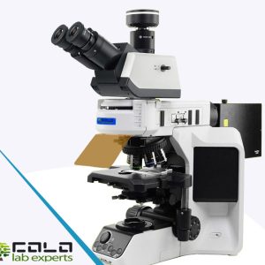
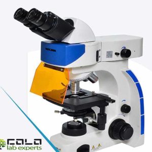
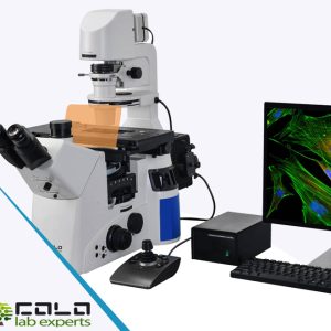
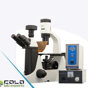
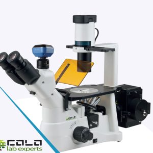
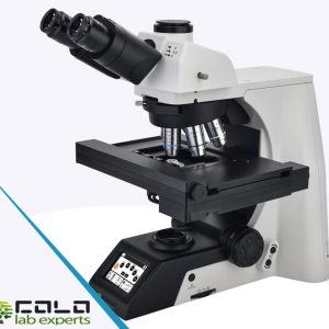
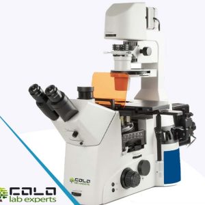
Featured: COLO.Science fluorescence systems
COLO.Science provides complete fluorescence imaging solutions, including brightfield-fluorescence microscopes, LED illumination, filter cubes, high-NA objectives and camera integration. We also offer installation, calibration and training through the LabServis partner network.
Fluorescence Microscopy Categories
Fluorescence Microscopy
Showing all 7 results