SOTLA500
Research Inverted Fluorescence Microscope · Binocular Imaging Platform
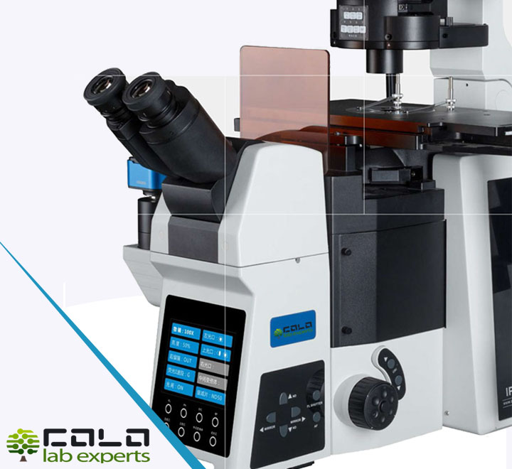
SOTLA500 is a research inverted fluorescence microscope designed for advanced transmitted light and fluorescence observation workflows. The platform combines infinity optics, a binocular head with tilt adjustment, a 6-position coded nosepiece, long working distance semi-apochromatic phase objectives, a motorized fluorescence cube holder, fine Z-axis control and PC-based imaging workflow for cell culture, fluorescence documentation and laboratory image analysis.
Inverted microscope
Fluorescence ready
Infinity optics
6-position nosepiece
Phase contrast
Motorized filter cubes
LED fluorescence source
USB 3.1 camera
PC control
Long working distance
Manual XY stage
Research applications
Product
SOTLA500 · research inverted fluorescence microscope
Address (HQ)
Polje ob Sotli 4
SI-3255, Slovenia
SI-3255, Slovenia
Contact
Document ID: COL-TEC-SOTLA500-EN
Version: Responsive layout
Informational technical sheet · Non-binding · Controlled electronic document
Version: Responsive layout
Informational technical sheet · Non-binding · Controlled electronic document
SOTLA500 Research Inverted Fluorescence Microscope Binocular head · fluorescence imaging · monochrome camera workflow
100/0 · 50/50 · 0/100
FN22 eyepieces
20–45° head tilt
IPD 50–76 mm
Stage 300 × 240 mm
Travel 135 × 85 mm
Condenser NA 0.55
8 filter cube capacity
400–645 nm LED
25,000 h source life
6.4 MP camera
USB 3.1
Technical specifications – Microscope platform
| Parameter | Specification / Value |
|---|---|
| Microscope type | SOTLA500 research inverted fluorescence microscope, binocular configuration |
| Optical system | Infinity optical system |
| Light path | 100/0 – 50/50 – 0/100 |
| Camera attachment | Left side camera attachment, 1× magnification |
| Focus shift range | Upper side: 6.5 mm or more from the starting position; bottom side: 3 mm or more from the starting position; initial position: 1 mm or more above the table surface |
| Head lighting source support | Lamp housing on top, tiltable up to 30° |
| Eyepieces | 10× eyepieces, one with diopter adjustment, field number FN22 |
| Binocular head | Binocular viewing head with tilting adjustment 20–45°, interpupillary distance 50–76 mm, eyepiece diopter range -5 to +5 |
| Stage | Manual stage size 300 mm (X) × 240 mm (Y); moving range 135 mm (X) × 85 mm (Y); stage thickness 30 mm |
| Stage controls | Right universal handle; X/Y axes limitable and lockable; 50 × 50 mm moving range with 110 mm replaceable disc |
| Condenser support | Up-and-down movement range of condenser support: 88 mm or more, with condenser centering mechanism |
| Condenser | NA 0.55, working distance 27 mm, dome with minimum 5 holes for adding optical elements |
| Phase contrast support | Optical elements for phase contrast corresponding to the offered lenses |
| Nosepiece | Manual coded nosepiece with 6 objective positions, revolving and reversed design |
| Brightfield illumination option | 10W LED light box (cold color temperature) or optional 12V / 100W halogen light box with preset filament center, depending on selected configuration |
Technical specifications – Objectives & fluorescence module
| Parameter | Specification / Value |
|---|---|
| Objective set | Infinity correction long working distance semi-apochromatic objectives with phase contrast support |
| Objective 1 | 10×, NA 0.30, WD 8.8 mm |
| Objective 2 | 20×, NA 0.45, WD 6.5–7.6 mm, coverslip thickness 0–2 mm |
| Objective 3 | 40×, NA 0.60, WD 2.85–4.05 mm, coverslip thickness 0–2 mm |
| Objective 4 | 60×, NA 0.70, WD 1.42–2.10 mm, coverslip thickness 0–1.3 mm |
| Fluorescent illuminator | Motorized filter cube holder with capacity for at least 8 filter cubes; simple tool-free cube installation |
| Fluorescence control | Motorization controlled via computer and manual steering wheel or joystick |
| Filter cube set 1 | Excitation filter 360–370 nm, emission filter 420–460 nm or wider ranges |
| Filter cube set 2 | Excitation filter 460–495 nm, emission filter 510–550 nm or wider ranges |
| Filter cube set 3 | Excitation filter 590–650 nm, emission filter 663–738 nm or wider ranges |
| Fluorescence light source | LED, emission spectrum 400–645 nm, internal fan cooling |
| Light intensity control | 0–100% in 1% steps, with manual control |
| Source life | More than 25,000 operating hours |
| Fine focus / Z-axis | Motorized fine focus (Z axis), with PC and joystick control |
Technical specifications – Camera & imaging software
| Parameter | Specification / Value |
|---|---|
| Camera type | 6.4 MP or higher monochrome camera |
| Sensor | Back-illuminated monochromatic CMOS |
| Sensor size | 1/1.8 inch (7.41 mm × 4.98 mm) or larger |
| Resolution | 3088 × 2076 pixels (photography) |
| Pixel size | 2.4 × 2.4 μm |
| Binning | 2 × 2 |
| Exposure range | 13 μs – 25 s |
| Live frame rate | Minimum 45 fps at 3088 × 2076 and up to 60 fps at minimum 1920 × 1080 resolution |
| Cooling | Passive cooling |
| Data transfer | USB 3.1 cable |
| Camera mount | C-mount type |
| Camera adapter | 0.5× magnification, C-mount type |
| Optional C-mount adapters | 0.5× / 0.65× / 1× C-mount adapters, focus adjustable |
| Included workflow elements | Electric control box, PC and monitor, centering eyepiece and professional software |
| Software functions | Image overlay, manual object counting, movie playback, side-by-side image comparison, snap and movie acquisition, 3D orthogonal plane section, time-lapse at specific intervals, automatic merging of photos from multiple fluorescence channels, Z-axis image joining, panoramic sample photography, geometry / combination / filter processing, area and line measurement, live image blur correction, 2D deconvolution, 3D deconvolution, confluency check and object tracking |
Key features of SOTLA500
Inverted research platform
- Research inverted fluorescence microscope layout for cell and culture observation
- Infinity optical design with binocular head and selectable light path
- Manual coded 6-position nosepiece for multiple objective configurations
- Long working distance semi-apochromatic objective setup
- Manual stage with large 300 × 240 mm platform and 135 × 85 mm travel
- Condenser centering and phase contrast support for advanced observation modes
Fluorescence & precision control
- Motorized filter cube holder with at least 8 cube capacity
- Tool-free filter cube installation for efficient workflow changes
- Fluorescence LED source with 400–645 nm emission spectrum
- Light intensity adjustment from 0 to 100% in 1% steps
- Motorized fine focus on Z axis with PC and joystick control
- Extended service life above 25,000 hours for the LED fluorescence source
Imaging & software workflow
- 6.4 MP or higher monochrome camera with back-illuminated CMOS sensor
- USB 3.1 data transfer for high-speed imaging workflow
- Time-lapse, multi-channel fluorescence merge and Z-axis joining
- Panoramic photography and comparative image review functions
- 2D and 3D deconvolution tools for image enhancement
- Measurement, object tracking and confluency evaluation support
Typical applications
- Live cell and cell culture fluorescence observation
- Phase contrast imaging of unstained biological samples
- Multi-channel fluorescence image documentation
- Laboratory research microscopy and academic applications
- Time-lapse imaging and sequence acquisition workflows
- Z-stack capture and extended focus image combination
- Panoramic sample stitching and comparative imaging
- Quantitative image analysis, counting and measurement
Configured package in this specification
| Configured microscope set | SOTLA500 microscope body, binocular head, 10× eyepieces, coded 6-position nosepiece, listed long working distance semi-apochromatic objective set, manual stage, condenser assembly and fluorescence module |
| Fluorescence bundle | Motorized filter cube holder with minimum 8 positions, three listed filter cube ranges and LED fluorescence light source with 400–645 nm emission spectrum |
| Imaging bundle | 6.4 MP or higher monochrome USB 3.1 camera, C-mount connection, 0.5× camera adapter, electric control box, PC, monitor, centering eyepiece and professional imaging software |
| Configuration note | The source text mentions both 10W LED and optional 12V / 100W halogen transmitted light options; final delivered configuration should follow quotation and order confirmation. |
Configuration note – SOTLA500
This technical sheet is arranged from the uploaded SOTLA500 technical catalog. The document combines microscope platform data, fluorescence module details and camera/software workflow. Some catalog lines appear abbreviated, so this sheet keeps the original technical meaning while organizing the content into the COLO.Science format. Final supply scope should always be confirmed in the quotation.
This technical sheet is arranged from the uploaded SOTLA500 technical catalog. The document combines microscope platform data, fluorescence module details and camera/software workflow. Some catalog lines appear abbreviated, so this sheet keeps the original technical meaning while organizing the content into the COLO.Science format. Final supply scope should always be confirmed in the quotation.
Frequently asked questions – SOTLA500
Is this an inverted microscope? Yes. SOTLA500 is specified as a research inverted fluorescence microscope.
Does it support fluorescence work? Yes. It uses a motorized fluorescence filter cube holder with at least 8 positions and an LED fluorescence source.
Can it be used for phase contrast? Yes. The condenser and objective description include phase contrast optical elements corresponding to the offered lenses.
Is digital imaging included? Yes. The specification includes a monochrome camera of 6.4 MP or higher with USB 3.1 transfer and C-mount connection.
Can it support advanced imaging software functions? Yes. The software list includes time-lapse, channel merging, Z-axis joining, panoramic imaging, deconvolution, tracking and measurement.
How is fluorescence control handled? The filter cube holder is motorized and controlled by computer as well as a manual steering wheel or joystick.
Please note: If this inverted fluorescence microscope configuration does not exactly match your application, contact our team. We can help with objective selection, fluorescence channel setup, camera choice, stage workflow and image analysis configuration. Specifications subject to change without notice.
Contact & Support
COLO LabExperts
Polje ob Sotli 4
SI-3255, Slovenia
Support note: Contact us for inverted microscope configuration, fluorescence filter selection, camera integration and microscopy workflow support.
Quick contact
Manufacturer & configuration notice:
Specifications in this document are based on the uploaded SOTLA500 technical catalog and arranged into the COLO.Science technical sheet format.
For procurement and tender purposes, the only valid and binding specification is the official COLO.Science quotation and the accompanying technical specification that is engineering-approved.